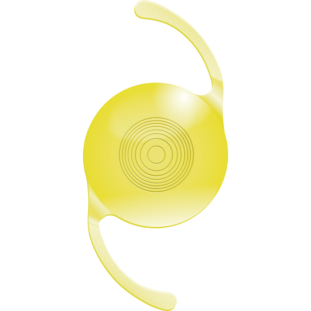
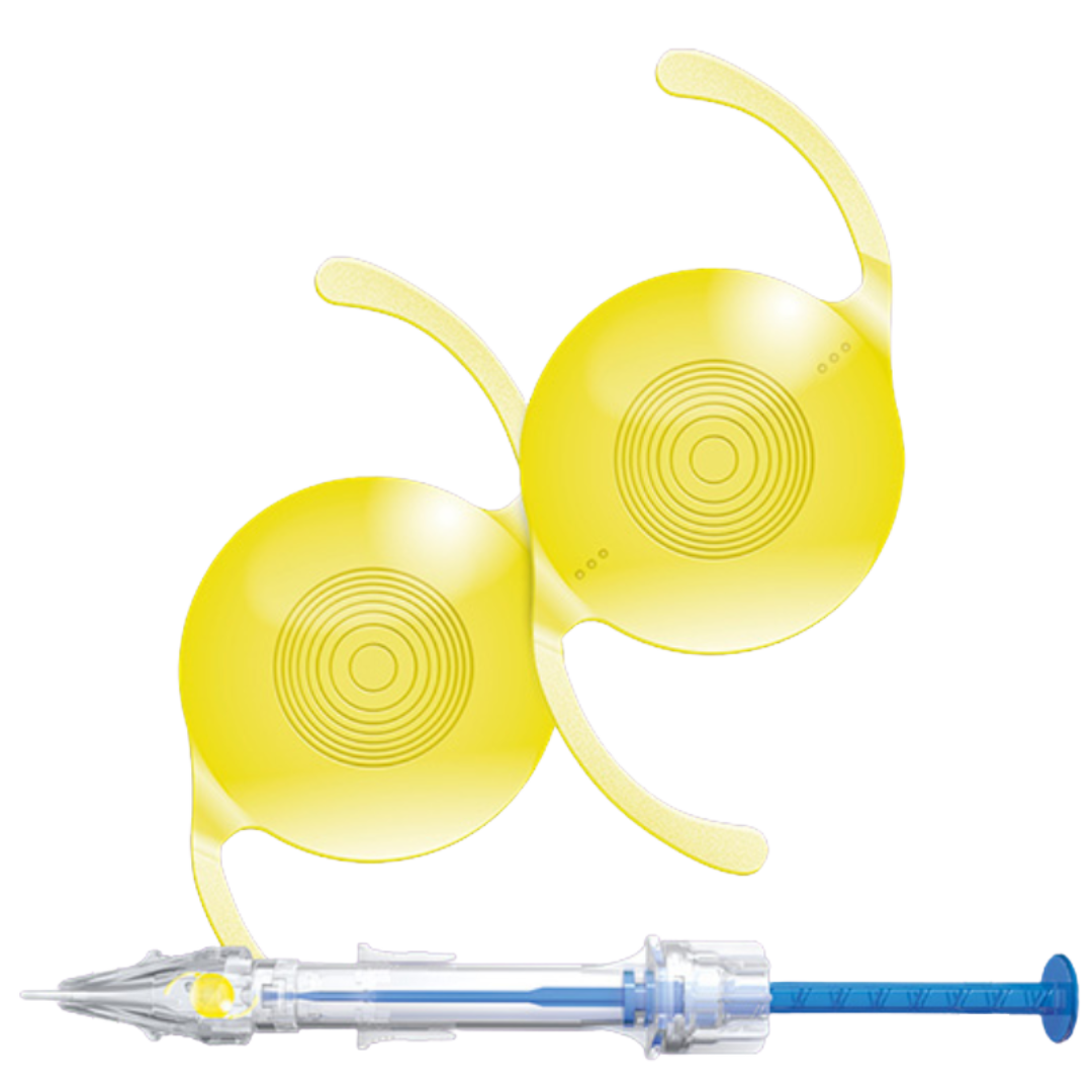
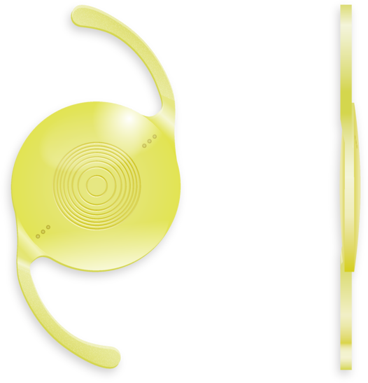
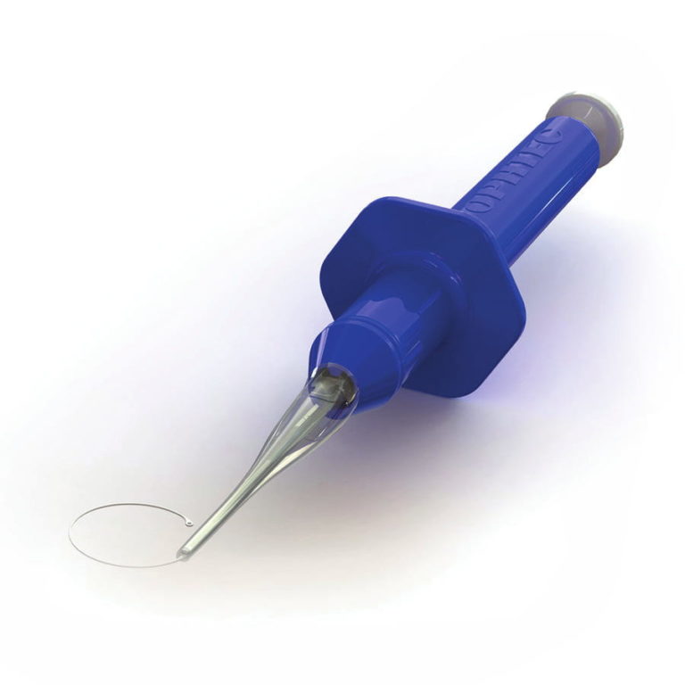
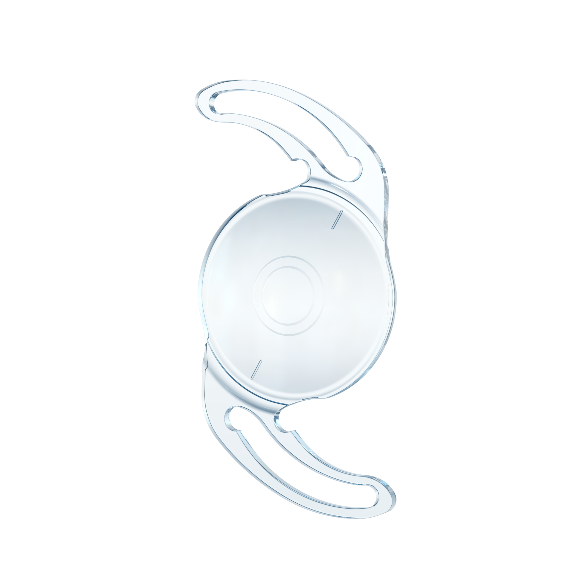
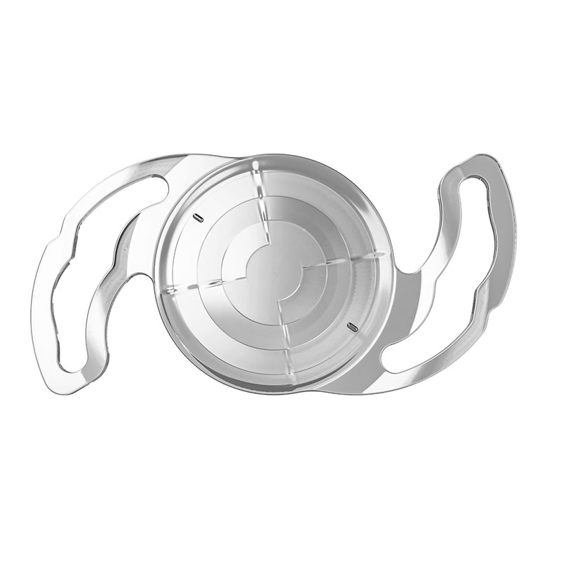
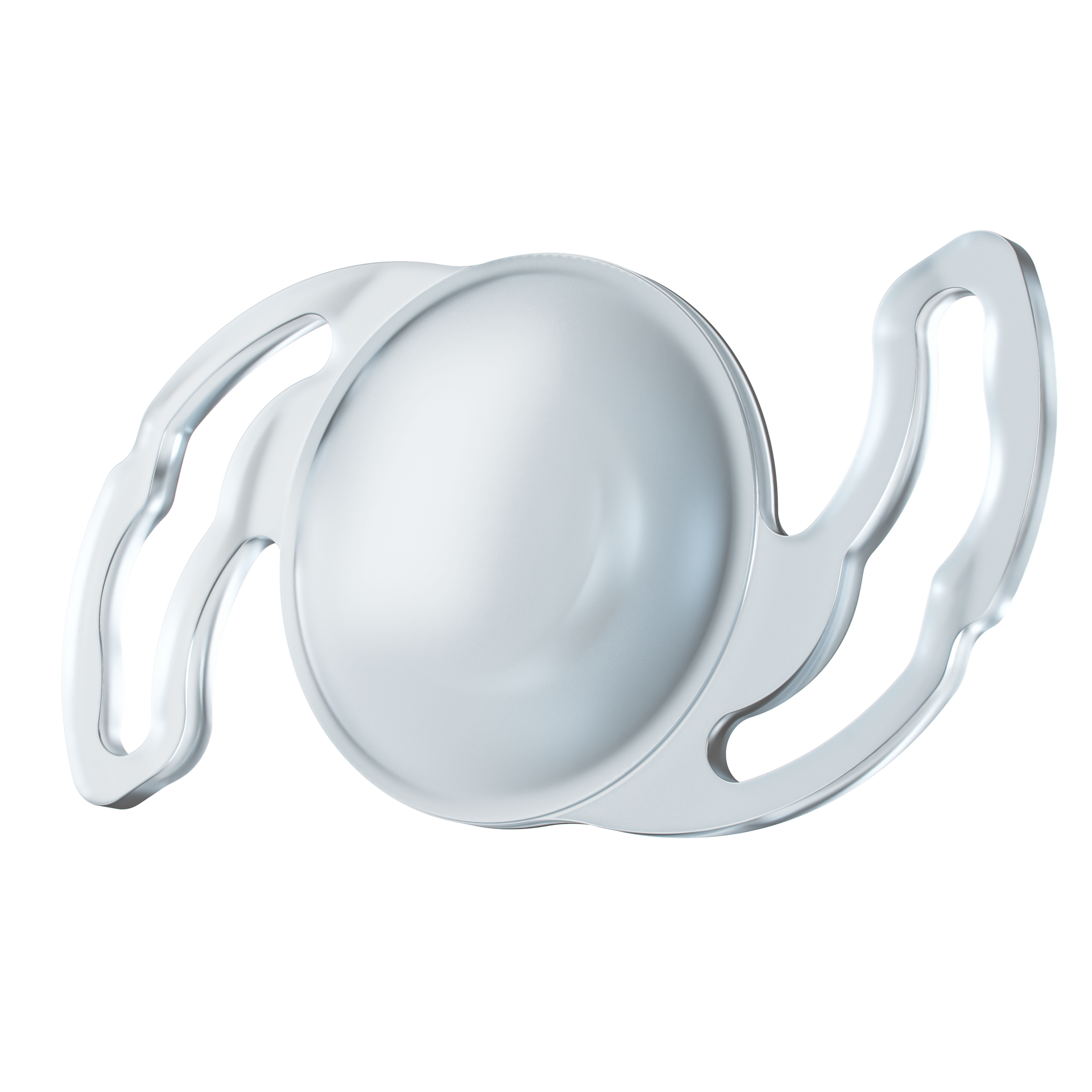
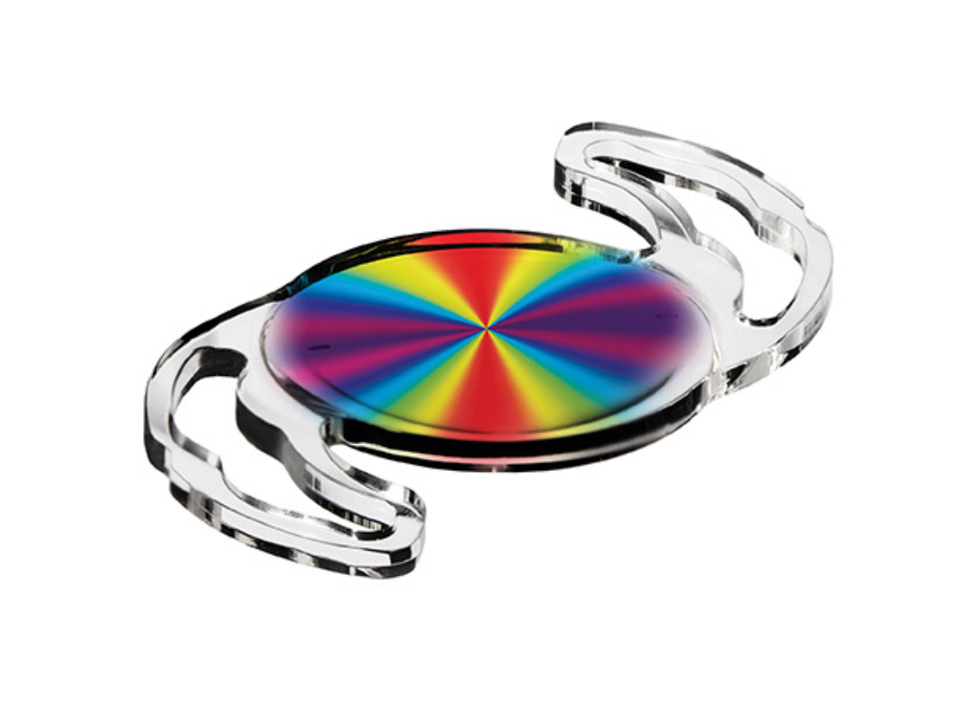
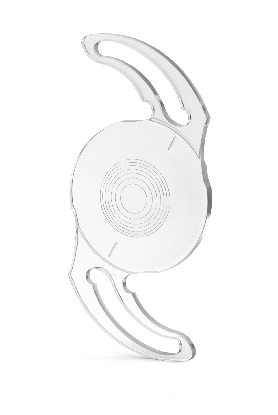
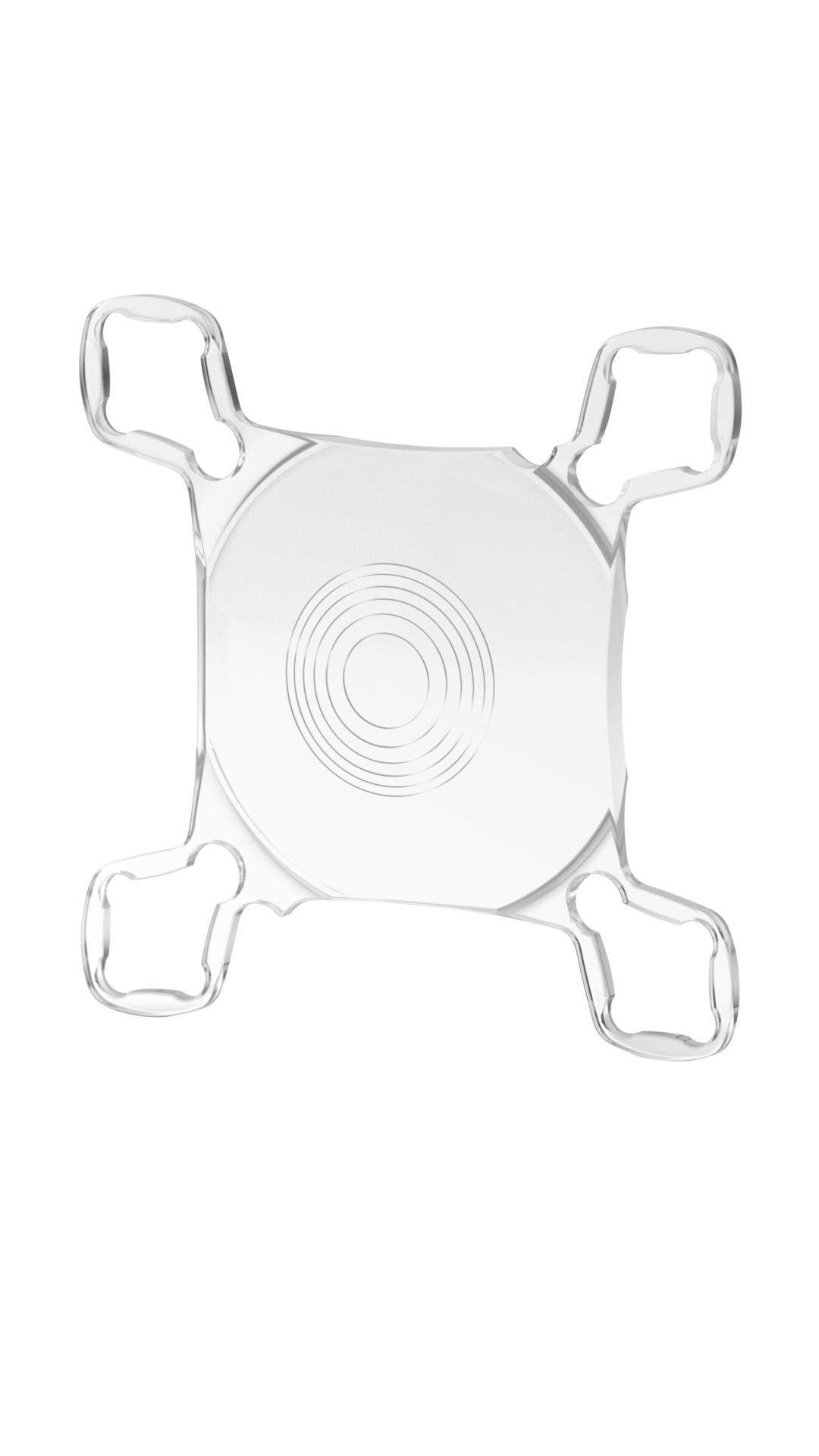
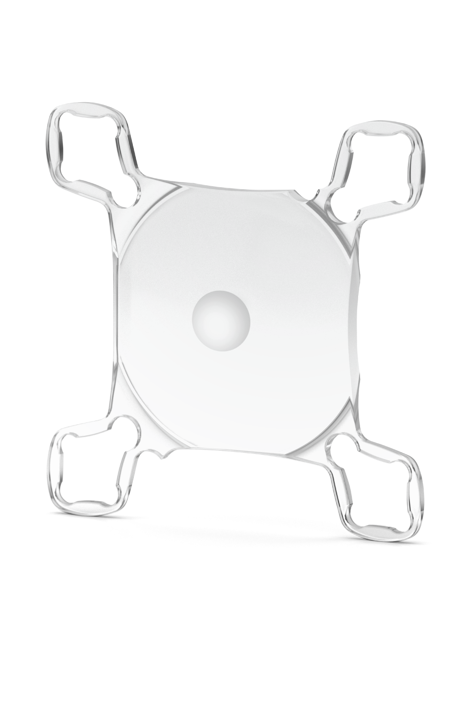
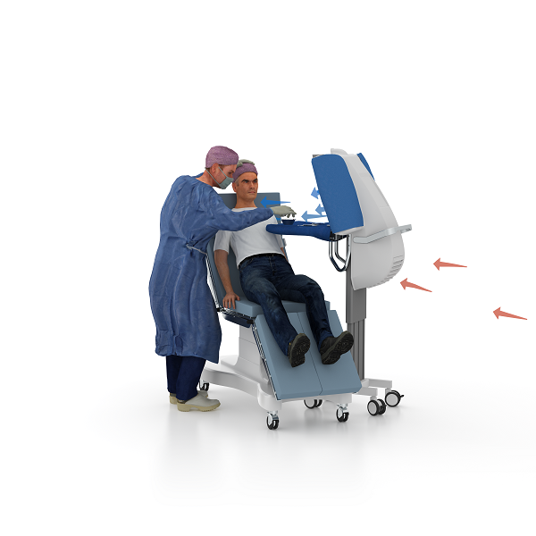
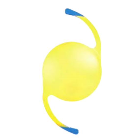
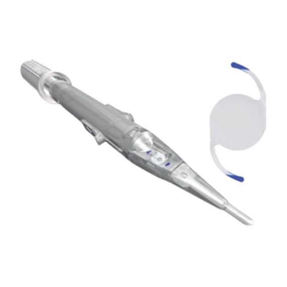
Optopol
Revo FC OCT
REVO FC the all-in-one high-resolution OCT with true colour fundus imaging and real-time hardware eye tracking function.
All-in-one OCT
Optopol REVO FC with cutting-edge colour fundus imaging for a new level of diagnostic certainty. High quality OCT scanning and a comprehensive analysis of the retinal layers combined with a fundus imaging make examinations more versatile than ever before.
Multi-use
An all-in-one device you can use in a number of ways:
- as a full colour Fundus Camera
- as a combo providing simultaneous OCT and fundus images
- for high quality OCT imaging including OCT-A
- as a Biometry device
Integrated non-mydriatic 12.3 Mpix Fundus Camera
- Advanced optical system for high quality imaging at a viewing angle as wide as 45°.
- Reduce the number of shots per eye and link a single fundus photo to several OCT exams.
- RGB channel, brightness, contrast, gamma and sharpness adjusters used with filters deliver a stunning retinal image.
- Detailed photos of a single or both eyes as well as a time comparison of fundus photos.
AccuTrack™
The REVO FC now comes with a real-time hardware eye tracking function which
compensates blinks, loss of fixation and involuntary eye movements during OCT
scanning.
AI Denoise™
Improved tomogram quality powered by Artificial Intelligence. Advanced AI algorithms enhance the quality of a single tomogram to the level of an averaged tomogram obtained through multiple scanning.
Full-range
Perfect for diagnosing even highly myopic patients, New Extended Depth™ retina imaging provides scans of increased depth for reliable and convenient observation of challenging cases.
Follow-up
High density of standard 3D scan allows the operator to precisely track disease progression. Analyse changes in morphology, quantified progression maps and evaluate the progression trends.
DICOM, EMR, network integration
DICOM connectivity allows the connection into large hospital medical systems. Send worklists (MWL) and reports (C-storage) or the whole examination to viewing stations. CMDL interface enables the integration of the REVO into practice management systems.
Key features:
- 3D Retina
- Biometry OCT
- OCT corneal topography
- Angio OCT
- Glaucoma analysis GCL and RNFL
- Anterior segment imaging
- Allows up to 3m between patient and clinician
Download
Technical specifications
Brochures & Manuals
Description & Technical Specs
FUNDUS CAMERA |
|
Type |
Non-mydriatic fundus camera |
Photography type |
Colour |
Angle of view |
45° ± 5% or less |
Min. pupil size for fundus |
3.3 mm or more |
Camera |
12.3 Megapixel CCD camera |
Camera modes |
|
Photography |
Fundus (Retina, Central, Disc, Manual fixation), Anterior photo, |
Flash adjustment, Gain, Exposure |
Auto, Manual |
Intensity levels |
High, Normal, Low |
OPTICAL COHERENCE TOMOGRAPHY |
|
Technology |
Spectral Domain OCT
|
Light Source |
SLED, Wavelength 850 nm |
Bandwidth |
50 nm half bandwidth |
Scanning speed |
80 000 measurements per second |
Axial resolution |
2.6 μm digital, 5 μm in tissue |
Transverse Resolution |
12 μm, typical 18 μm |
Overall scan depth |
2.8 mm / ~6 mm in Full Range mode |
Min. pupil size for OCT |
2.4 mm |
Focus adjustment range |
-25 D to +25 D |
Scan range |
Posterior 5 mm to 15 mm, Angio 3 mm to 9 mm, Anterior 3 mm to 18 mm |
Scan types |
3D, Angio*, Full Range Radial, Full Range B-scan, Radial (HD), B-scan (HD), Raster (HD), Cross (HD), TOPO, AL, ACD |
Fundus alignment |
IR, Live Fundus Reconstruction |
Alignment method |
Fully automatic, Automatic, Manual |
Fundus Tracking |
Real time active, iTracking |
Retina analysis |
Retina thickness, Inner Retinal thickness, Outer Retinal thickness, RNFL+GCL+IPL thickness, GCL+IPL thickness, RNFL thickness, RPE deformation, MZ/EZ-RPE thickness |
Angiography OCTan optional software module to purchase |
Vitreous, Retina, Choroid, Superfi cial Plexus, RPCP, Deep Plexus, Outer Retina, Choriocapilaries, Depth Coded, SVC, DVC, ICP, DCP, Custom, Enface, FAZ, VFA, NFA, Quantifi cation: Vessel Area Density, Skeleton Area Density, Thickness map |
Glaucoma analysis |
RNFL, ONH morphology, DDLS, OU and Hemisphere asymmetry, Ganglion analysis as RNFL+GCL+IP and GCL+IPL, Structure + Function via connection with PTS software version 3.4 or higher |
Angiography mosaic |
Acquistion method: Auto, Manual Mosaic modes: 10 x 6 mm, Manual up to 12 images |
Biometry OCTan optional software module to purchase |
AL, CCT, ACD, LT, P, WTW IOL Formulas: Hoffer Q, Holladay I, Haigis, Theoretical T, Regression II |
Corneal Topography Mapan optional software module to purchase |
Axial [Anterior, Posterior], Refractive Power [Kerato, Anterior, Posterior, Total], Net Map, Axial True Net, Equivalent Keratometer, Elevation [Anterior, Posterior], Height, KPI (Keratoconus Prediction Index) |
AnteriorNo adapter required |
Anterior Chamber Radial, Anterior Chamber B-scan, Pachymetry, Epithelium map, Stroma map, Angle Assessment, AIOP, AOD 500/750, TISA 500/750, Angle to Angle view, Wide Angle, Wide Cornea |
Connectivity |
DICOM Storage SCU, DICOM MWL SCU, CMDL, Networking |
Fixation target |
OLED display (The target shape and position can be changed), External fixation arm |
Dimensions (LxWxH) / Weight |
479 × 367 × 493 mm / 30 kg |
Power supply / consumption |
100-240 V, 50/60 Hz / 90-110 VA |